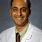
We are pleased to announce that Dr. Neil Sanghvi has joined First Coast Heart & Vascular Center. Dr. Sanghvi has been recognized as a leader in the field of clinical electrophysiology and is honored to carry the distinction of being a Fellow of the Heart Rhythm Society as well as a Fellow of the American College of Cardiology. His current interests include improving techniques in atrial fibrillation ablation, minimally invasive closure of the left atrial appendage for stroke prevention, and management of congestive heart failure with biventricular devices. Dr. Sanghvi actively participates in national clinical research trials and has authored several papers on the topic of arrhythmia management.
“I believe that each patient deserves attention, compassion, respect, and the opportunity to understand their particular ailment. I strive to provide personalized care and take pride in being able to clearly communicate with my patients. My reward is seeing a patient
successfully and safely complete a procedure and witness the often immediate improvement in their quality of life.”
Dr. Sanghvi began his interested with medicine early in high-school. He was accepted and attended the prestigious 7-year medical program at Boston University. From there, he returned to his home state of New Jersey where he completed medical school at Rutgers- New Jersey Medical School. Dr. Sanghvi found that he missed Boston, so he returned to Boston University for his Internal Medicine Residency. He then travelled to Washington,D.C. to participate in the George Washington University Cardiology Fellowship Program. He completed his Electrophysiology training at the prestigious Weill Cornell Medical Center in New York City. Dr. Sanghvi practiced for 5 -years in New York City before arriving to Florida. He was instrumental in expanding the complex ablation program at Lenox Hill Hospital in Manhattan.
Common disorders that Dr. Sanghvi manages include atrial fibrillation, SVT,ventricular tachycardia, syncope (passing out), palpitations, pacemakers, and ICDs (defibrillators).
According to the Centers for Disease Control and Prevention (CDC), 60% of Americans are not meeting the recommended levels of physical activity. Fully 16% of Americans are not active at all. Overall, women tend to be less active than men, and older people are less likely to get regular physical activity than younger individuals.
In a recent article published in Circulation: Journal of the American Heart Association, researchers from the Harvard Medical School released data from the 10-year Women’s Health study that showed moderate exercise reduced the risk of heart disease by 27% to 41%. the study was performed on 27,055 participants.
The mechanism of benefit was shown to be largely due to the reduction of LDL (bad cholesterol), raising HDL (good cholesterol) and reducing inflammation.
It is well-known that inflammation leads to the release of molecules called cytokines that can cause damage to the blood vessels in the heart and throughout the body. Cholesterol tends to deposit at these damaged sites, leading to plaque. the damaged vessels containing increased cholesterol deposits are the sites where platelets attach to blood vessels. These clumps of platelets can break off and completely block the blood vessel as it narrows downstream. In the coronary arteries, this leads to an acute heart attack; in the brain, it causes either a major or a minor stroke.
As little as two hours of brisk walking every week was sufficient to lower the risk of major cardiac events dramatically. Can you imagine that if all you do is walk briskly for 17 minutes daily, your risk of heart disease will go down by 40%? Regular physical activity is defined as about 30 minutes of moderate activity (preferably all days of the week) can reduce the risk of heart disease. One can lower your chances of having a stroke, colon cancer, high blood pressure, diabetes and other medical problems.
If you’re also trying to manage your weight and prevent gradual, unhealthy weight gain, try to get 60 minutes of moderate to vigorous-intensity activity on most days of the week. At the same time, watch your calorie intake. Take in only enough calories to maintain your weight. I often counsel my patients to eat no more than 350 calories a meal four to six times a day. Most of our bodies cannot metabolize more than this amount so large meals that many of us are used to will cause us to store weight and develop fatty tissue.
I often share the following scenario with my patients:
Patient A – Eats two large meals daily of 750 calories each – totalling 1500 calories.
Patient B – Eats five small meals of 300 calories each about 2-3 hours apart – totalling 1500 calories.
Q: Who will lose weight?
A: Patient B
By eating only the calories the body can burn, patient B will likely lose weight coupled with regular physical activity. Unfortunately, Patient A is unable to burn the extra 450 calories they eat at each meal and that totals 900 extra calories daily. 1 pound is about 3500 calories. In about 4 days, a person can gain an unwanted pound of weight eating this way and it can be exacerbated if they are physically inactive.
Another example: A 200 pound person who keeps on eating the same amount of calories, but walks briskly each day for 1.5 miles, will lose about 14 pounds in one year. Staying active will also help to keep the weight off. Second, you can eat fewer calories and be more active. This is the best way to lose weight, since you’re more likely to be successful by combining a healthful, lower-calorie diet with physical activity.
For example, a 200 pound person who consumes 250 fewer calories per day, and begins to walk briskly for 1.5 miles each day will lose about 40 pounds in one year.
As you can see, about 2/3 of weight loss is attributed to diet, but we need physical activity to help us stave off medical problems including heart disease.
Heart Disease
Heart disease occurs when the arteries that supply blood to the heart muscle become hardened and narrowed, due to a buildup of plaque on the arteries’ inner walls. Plaque is the accumulation of fat, cholesterol and other substances. As plaque continues to build up in the arteries, blood flow to the heart is reduced. Heart disease can lead to a heart attack. A heart attack happens when a cholesterol-rich plaque bursts and releases its contents into the bloodstream. This causes a blood clot to form over the plaque, totally blocking blood flow through the artery and preventing vital oxygen and nutrients from getting to the heart. A heart attack can cause permanent damage to the heart muscle. Some people aren’t too concerned about heart disease because they think it can be cured with surgery.
This is a myth. Heart disease is a lifelong condition. It’s true that certain procedures can help blood and oxygen flow more easily to the heart. But the arteries remain damaged, which means you are still more likely to have a heart attack. What’s more, the condition of you blood vessels will steadily worsen unless you make changes in your daily habits and control other factors that increase risk.
You have control.
Physical inactivity is one of several major risk factors for heart disease that you can do something about. The others are:
Smoking. People who smoke are up to six times more likely to suffer a heart attack than non-smokers, and the risk increases with the number of cigarettes smoked each day.
High Blood Pressure. Also known as hypertension, high blood pressure increases your risk of heart disease, stroke, kidney disease, and congestive heart failure.
High Blood Cholesterol. High Blood Cholesterol can lead to the buildup of plaque in your arteries, which raises the risk of a heart attack. Starting at age 20, everyone should have their cholesterol levels checked by means of a blood test called a “lipoprotein profile”. You can lower high blood cholesterol by getting regular physical activity, eating less saturated fat and trans fat, and managing your weight. In some cases, medication is also needed.
Overweight. If you are overweight or obese, you are more likely to develop heart disease even if you have no other risk factors. Ask your doctor to help you determine whether you need to lose weight for your health. The good news: Losing just 5-10% of your current weight will help to lower your risk of heart disease and many other medical disorders.
Diabetes greatly increases your risk for heart disease, stroke, and other serious diseases. Ask your doctor whether you should be tested for it. Many people at high risk for diabetes can prevent or delay the disease by reducing calories as part of a healthy eating plan, and by becoming more physically active.
Taking Precautions
Some people should get medical advice before starting, or significantly increasing physical activity. Check with your doctor first if you:
Are over 40 years old and not used to moderately energetic activity.
Currently have a heart condition, have developed chest pain within the last month, or have had a heart attack. (Also see the section, “After a Heart Attack”)
Have a parent or sibling who developed heart disease at an early age.
Have any other chronic health problem or risk factors for a chronic disease.
Tend to easily lose your balance or become dizzy.
Feel extremely breathless after mild exertion.
Are on any type of medication.
While physical activity can strengthen the heart, some types of activity may worsen existing heart problems. Warning signals include sudden dizziness, cold sweat, paleness, fainting, extreme breathlessness, or pain or pressure in your upper body. These symptoms may occur during, or just after, an activity. Ignoring these signals and continuing your activity may lead to serious heart problems. Instead, call your doctor right away.
Overall, it is best to get regular comprehensive evaluations to help understand how best to incorporate the right type of exercies in your life to help reduce your risk of heart disease and other illnesses.
We at First Coast Heart & Vascular Center are delighted to welcome you to our new practice and sincerely appreciate the opportunity to care of you and your family’s heart and vascular needs.
We would like to remind you of the annual open enrollment season for Medicare participants. Many insurance carriers are offered and we take ALL of them with the exception of Humana Gold and Humana Gold Plus.
First Coast Heart & Vascular Center and all its providers are pleased to inform you of our continued participation with United HealthCare – Medicare AARP and we are in network.
We are growing and would love for you to be part of our family as our growth continues. First Coast Heart & Vascular Center is committed to providing quality ,efficient, compassionate and cost effective care. If you have further questions, please do not hesitate to contact our practice.
We appreciate your trust and confidence in our practice. We wish all of you a happy and safe holiday season.
Atrial Fibrillation – also known as AFib or AF – is the most common arrhythmia. It affects more than 2.5 million American adults and 4.5 million people living in the European Union, and accounts for approximately one-third of hospitalizations for cardiac rhythm disturbances.
It is characterized by rapid and irregular heartbeat caused when the top chambers of the heart (the atria) quiver (fibrillate) erratically, sometimes faster than 20o times per minute.
AFib can also increase the risk of stroke fivefold. It is estimated to be responsible for 88,000 deaths and $16 Billion in additional costs to the U.S. Healthcare system. As the world population ages, the prevalence of AFib is projected to increase. In fact, in the next 30-40 years, the number of people diagnosed with AFib in the U.S. is expected to more than double.
Here’s how patients have described their experience:
“My heart flip-flops, skips beats, and feels like its banging against my chest wall, especially if I’m carrying stuff up my stairs or bending down.”
What happens during AF?
Atrial Fibrillation (AF) is the most common type of irregular heartbeat. Normally, you heart contracts and relaxes to a regular beat. In atrial fibrillation, the upper chambers of the heart (the atria) beat irregularly (quiver) instead of beating effectively to move blood into the ventricles. About 15-20 percent of people who have strokes have arrhythmia.
Additional common symptoms of atrial fibrillation
Sometimes people with AF have no symptoms and their condition is only detectable upon physical examination. Still, others may experience one or more of the following symptoms:
Rapid and irregular heartbeat
Fluttering or “thumping” in the chest
Dizziness
Shortness of breath and anxiety
Weakness
Faintness or confusion
Fatigue when exercising
Sweating
*Chest pain or pressure
*Chest pain or pressure is a medical emergency. You may be having a heart attack. Call 9-1-1 immediately.
Are there different types of AF? Do they have different symptoms?
The symptoms are generally the same; however the duration of the AF and underlying reasons for the condition help medical practitioners classify the types of AF problems.
Paroxysmal fibrillation is when the heart returns to a normal rhythm on its own. People who have this type of AF may have episodes only a few times a year or their symptoms may occur every day. These symptoms are very unpredictable and often can turn into a permanent form of atrial fibrillation
Persistent AF is defined as an irregular rhythm that lasts for longer than 7 days. This type of AF will not return to normal sinus rhythm on its own and will require some form of treatment.
Permanent AF occurs when the condition lasts indefinitely and can no longer be controlled with medication.
Over a period of time, paroxysmal fibrillation may become more frequent and longer lasting, sometimes leading to permanent or chronic AF. All types of AF can increase your risk of stroke. Even if you have no symptoms at all, you are nearly 5 times more likely to have a stroke than someone that does not have atrialfibrillation.
Know your treatment goals
The treatment goals of atrial fibrillation (AF) start with a proper diagnosis through an in-depth examination from a physician. The exam usually includes questions about your history and often an EKG or ECG. Some patients may need a thorough electrophysiology study.
Prevention and Risk Reduction
After a patient is diagnosed with atrial fibrillation, the ideal goals may include:
Restoring the heart to a normal rhythm
Reducing an overly high heart rate
Preventing blood clots
Managing risk factors for stroke
Preventing additional heart rhythm problems
Preventing heart failure
1. How will I prevent stroke?
Depending on your risk, you will likely either need aspirin or warfarin or another type of anticoagulation medication.
2.Are there additional lifestyle modifications important for stroke prevention?
3.What is my C.H.A.D.S. risk? Congestive heart failure Hypertension Age (75 or greater) Diabetes Stroke (prior episode)
4. Are there options to control my heart rate and this irregular heart rhythm?
Based on your past medical history and risk for having a future stroke, there are several options for you and your health care provider to discuss in order to manage your AF.
Medications for Atrial Fibrillation (AF)
Medications are often prescribed to prevent and treat blood clot which can lead to a stroke. The longer you have untreated AF, the less likely it is that normal rhythm can be reestablished.
Medication options may include blood thinners, rate controllers, and rhythm controllers. Lists included here are not intended to be comprehensive, and we encourage you to contact our office to keep up with the newest in AF medication options.
Preventing Clots with Medication (antiplatelets and anticoagulants)
Drugs such as blood thinners are given to patients to prevent blood clot formation or to treat an existing blood clot. Examples include Aspirin, Warfarin and other FDA Approved anticoagulants.
Important Precautions when taking anti-clotting medications
Call your healthcare provider right away if you have any unusual bleeding or bruising
If you forget to take your daily anticoagulant dose, Don’t take an extra one to catch up! Follow your healthcare provider’s directions about what to do if you miss a dose.
Always tell your doctor, dentist and pharmacist that you take one of these medicines.
Many drugs change the effects of these agents on the body. Even vitamins (and some foods) could change this effect.
Heart Rate Controlling Medication
Beta blockers, Calcium blockers and Digoxin are drugs used to slow the heart rate. Most people can function and feel better if their heart rate is controlled. Some examples may include Carvedilol, Metoprolol, Atenolol, Dilitazem, and Verapamil.
Heart Rhythm Controlling Medications
Once your heart rate is under control, the next management consideration is usually treating the abnormal heart rhythm with medications to restore the heart rhythm to normal. Your healthcare provider will most likely want to monitor progress closely. Examples include Flecainide (Tambocor), Propafenone (Rhythmol), Amiodarone, Carodarone or Pacerone, Sotalol (Betapace), or Dronedarone (Multaq).
The decision to use electrical cardio version
Your provider may recommend a trans esophageal Echocardiogram (TEE) as a first step. The TEE procedure involves swallowing a small ultrasound device that allows the healthcare team to look inside your heart atria for blood clots.
Radiofrequency Ablation or Catheter Ablation
Catheter ablationis an atrial fibrillation treatment that is done by a specialized cardiologist, called an Electro physiologist (EP) who deals with irregular heartbeats (arrhythmias).
It is a minimally-invasive procedure that is generally less invasive than surgery. It is a commonly-used treatment for atrial fibrillation as well as other cardiac arrhythmia. Like other atrial fibrillation treatments, it is most successful in treating paroxysmal atrial fibrillation, but much progress has been made in treating persistent and long standing persistent as well.
Ablation is used for cardiac arrhythmias when long-term medications or electrical cardio version are either not preferred or were not effective. Or when the patient prefers no to take any medications. Before ablation surgery, electrical mapping of the heart is performed. An electrically sensitive catheter is used to map the heart muscle and the origins of the “extra” electrical activity throughout the heart. The map tells the physician which areas of the heart are creating problematic electric signals that interfere with the proper rhythm. After a single procedure, more than 60-70% of patients with an otherwise normal heart can enjoy freedom from arrhythmias according to studies that have followed patients typically for one year. With two or more procedures, the efficacy can be as high as 80-90% in other recent case studies.
Catheter ablation is the only cardiac procedure that can be correctly called curative (No, stents are not curative)
How is an ablation performed?
A catheter (thin, flexible tube) is inserted into the patient’s blood vessels and is gently guided to the heart. The physician carefully destroys malfunctioning tissue using the catheter to deliver energy (such as radiofrequency, laser or cryotherapy) to scar the problematic areas. The scarred areas will no longer send abnormal signals. If successful, the heart will return to a normal rhythm. This minimally invasive procedure usually has a short recovery period. Patients are generally placed on a short course of anti-arrhythmic drugs while the procedure takes full effect.
Common types of Ablation include:
Pulmonary vein isolation ablation (PVI ablation or PVA) In some AF patients, fibrillation is triggered by extra electrical currents in the pulmonary veins. During this procedure, the catheter tip is used to destroy the tissue that is sending the extra currents and, in most cases, normal heart rhythm returns.
AV node ablation with pacemakers. In other AF patients, the trigger for their AF occurs in the AV node (the place where the electrical signals pass from the atria to the ventricles). The catheter is placed near the AV nod and a small area of tissue is destroyed. A pacemaker is then implanted to restore and maintain the heart’s normal rhythm.
At First Coast Heart & Vascular Center, our group treats atrial fibrillation conservatively with medical treatment, however for select cases our board certified electrophysiologist, Dr. Dinesh Pubbi can perform Atrial Fibrillation ablations at the hospitals that have state of the art EP labs.
Dr. Van Crisco’s interview with First Coast Living. Dr. Crisco discusses heart disease and interventional cardiology treatments. Cardiac Catheterization, Coronary Intervention, TAVR, Endovascular Intervention, Limb Salvage and other treatments.
First Coast Heart & Vascular Center Earns ACR Ultrasound Accreditation
First Coast Heart & Vascular Center has been awarded a three-year term of accreditation in ultrasound as the result of an extensive review by the American College of Radiology (ACR). Ultrasound imaging, also known as sonography, uses high-frequency sound waves to produce images of internal body parts to help providers diagnose illness, injury, or other medical problems.
The ACR gold seal of accreditation represents the highest level of image quality and patient safety. It is awarded only to facilities meeting ACR Practice Guidelines and Technical Standards, following a peer-review evaluation by board-certified physicians and medical physicists who are experts in the field. Image quality, personnel qualifications, adequacy of facility equipment, quality control procedures, and quality assurance programs are assessed. The findings are reported to the ACR Committee on Accreditation, which subsequently provides the practice with a comprehensive report they can use for continuous practice improvement.
The ACR is a national professional organization serving more than 36,000 diagnostic/interventional radiologists, radiation oncologists, nuclear medicine physicians, and medical physicists with programs focusing on the practice of medical imaging and radiation oncology and the delivery of comprehensive health care services.
P.A.D. is short for Peripheral Arterial Disease. People have P.A.D. when the arteries in their legs become narrowed or clogged with fatty deposits, or plaque. The buildup of plaque causes the arteries to harden and narrow, which is called atherosclerosis . When leg arteries are hardened and clogged, blood flow to the legs and feet is reduced. Some people call this poor circulation.
P.A.D. occurs most often in the arteries in the legs, but it also can affect other arteries that carry blood outside the heart. This includes arteries that go to the aorta, the brain, the arms, the kidneys and the stomach. When arteries that supply the heart are hardened or narrowed, it is called coronary artery disease or cardiovascular disease.
The good news is that like other diseases related to the arteries, P.A.D. can be treated by making lifestyle changes, by taking medicines, or by undergoing leg artery angioplasty or vascular surgery, if needed. And you can live well with P.A.D.
Is P.A.D. serious?
P.A.D. is a serious disease commonly affecting Americans over the age of 50. The hardened arteries found in people with P.A.D. are a sign that they are likely to have hardened and narrowed arteries to the heart and the brain. That is why people with P.A.D. have a two- to six-times greater chance of death from a heart attack or a stroke.
When the blood flow to the legs is greatly (or severely) reduced, people with P.A.D. may have pain when walking. P.A.D. may cause other problems that can lead to amputation of a toe, foot or a leg. People with P.A.D. may become disabled and not be able to go to work. As time goes on, they may have a very poor quality of life.
Who is at risk for P.A.D.?
The chance of having P.A.D. increases as you get older. People over age 50 have a higher risk for P.A.D., but the risk
is increased if you:
Smoke, or used to smoke
Have diabetes
Have high blood pressure
Have abnormal blood cholesterol levels
Are of African American ethnicity
Have had heart disease, a heart attack or a stroke
Have a family history of P.A.D., heart attack or stroke.
What are the warning signs or symptoms of P.A.D.?
P.A.D. develops slowly over many years. In the early stages, most people with P.A.D. have no symptoms. Only about one out of four people with P.A.D. actually feel typical symptoms of P.A.D. in their leg muscles (known as “claudication”, defined below). By that time, their arteries may be so clogged or hardened that they are not getting enough oxygen to supply their leg muscles.
The most common signs of P.A.D. include one or more of these problems:
Cramps, tiredness or pain in your legs, thighs or buttocks that always happens when you walk but that goes away when you rest. This is called claudication.
Foot or toe pain at rest that often disturbs your sleep.
Skin wounds or ulcers on your feet or toes that are slow to heal, or that do not heal.
Sometimes, people ignore their leg pain and think it is just a sign that they are getting older. As a result, many people with P.A.D. do not know they have it and do not get treatment. It is important to discuss any leg or thigh pain you may be having with your health care provider since it may be a warning sign of a serious disease such as P.A.D.
How do I find out if I have P.A.D.?
If you think you have P.A.D., see your health care provider and talk about any symptoms you are having and go over your medical history and your risk factors for P.A.D. Your provider will examine the pulses in your feet and legs. If your provider finds those pulses are weak and thinks you may have P.A.D., your provider may order a test called the ABI, which stands for ankle-brachial index .
The ABI is the best test for finding out if you have P.A.D. It uses sound waves to find out if there is reduced blood flow in the arteries. It also compares the blood pressure in your ankles with the blood pressure in your arms. P.A.D. also can be diagnosed by other tests that measure blood pressures in the leg (segmental pressure), toe pressures (toe-brachial index or TBI) or artery blood flow (with ultrasound). Other more sophisticated tests can be performed such as PVR ( pulse volume recordings), CTA ( Cat scan) , magnetic resonance angiography (MRA) or catheterization or angiography.
How is P.A.D. treated?
P.A.D. can be treated with lifestyle changes, medicines and surgery, if needed. Since people with P.A.D. are at high risk for heart attacks and stroke, they must take charge of controlling their risk factors related to cardiovascular disease.
These life-saving steps will help to prevent and control P.A.D.:
Get help to quit smoking and set a quit date now.
Lower your blood pressure to less than 140/90 mmHg or less than 130/80 mmHg if you have diabetes or chronic kidney disease.
Lower your LDL (bad) cholesterol to less than 100 mg/dl or to less than 70 mg/dl if you are at very high risk for a heart attack or stroke (if you smoke, have diabetes or have chronic kidney disease).
Manage your blood glucose to reach an A1C level of less than 7 and practice proper foot care if you have diabetes.
Talk to your doctor about taking antiplatelet medicines such as aspirin or clopidogrel (Plavix) to prevent clotting.
Follow a healthy eating plan to control your blood pressure, cholesterol and blood glucose (for diabetes).
Get regular exercise such as walking for 30 minutes at least 3 or 4 times per week.
If you have pain or cramps in your legs, ask your health care provider about an exercise program that will help improve your symptoms. If possible, get a referral to a special P.A.D. exercise program.
For most people with P.A.D., these life-saving steps may be enough to slow down the disease and even improve any symptoms. If needed, your health care provider can refer you to a specialist for procedures or surgery to treat arteries that are severely blocked. These procedures often help people with P.A.D. to improve symptoms and to avoid losing a foot or leg.
Remember:
Finding and treating P.A.D. early can help keep your legs healthy, lower your risk for heart attack or stroke, and save your life and limbs.
Our board certified interventional cardiologists and vascular specialists have an extensive experience with interventions to treat simple to complex vascular blockages in any vascular bed or organ including carotids ( neck arteries), arms , kidney arteries, stomach or intestinal arteries, leg arteries as well as heart arteries. Our experts use simple balloon angioplasty, stents to more complex atherectomy devices, laser catheters, and clot buster catheters to remove clots or cholesterol plaques. If you wish to make an appointment with our doctors, please contact us at (904)-423-0010 or visit our web site (www.firstcoastheart.com) for our 8 locations or phone numbers in three counties.
An abdominal aortic aneurysm, also called AAA or triple A, is a bulging, weakened area in the wall of the aorta resulting in an abnormal widening or ballooning greater than 50 percent of the vessel’s normal diameter (width). The aorta extends upward from the top of the left ventricle of the heart in the chest area (ascending thoracic aorta), then curves like a candy cane (aortic arch) downward through the chest area (descending thoracic aorta) into the abdomen (abdominal aorta). The aorta delivers oxygenated blood pumped from the heart to the rest of the body. The most common location of arterial aneurysm formation is the abdominal aorta, specifically, the segment of the abdominal aorta below the kidneys, or an infrarenal aneurysm. An aneurysm can be characterized by its location, shape, and cause.
The shape of an aneurysm is described as being fusiform or saccular, which helps to identify a true aneurysm. The more common fusiform-shaped aneurysm bulges or balloons out on all sides of the aorta. A saccular-shaped aneurysm bulges or balloons out only on one side.
The aorta is under constant pressure as blood is ejected from the heart. With each heart beat, the walls of the aorta distend (expand) and then recoil (spring back), exerting continual pressure or stress on the already weakened aneurysm wall. Therefore, there is a potential for rupture (bursting) or dissection (separation of the layers of the aortic wall) of the aorta, which may cause life-threatening hemorrhage (uncontrolled bleeding) and, potentially, death. The larger the aneurysm becomes, the greater the risk of rupture. Because an aneurysm may continue to increase in size, along with progressive weakening of the artery wall, surgical or endovascular intervention may be needed. Preventing rupture of an aneurysm is one of the goals of therapy.
It is estimated 1 to 4 % of persons over age 50 years are affected. Rupture of an AAA is the presenting feature of this disease in roughly 2/3rds of patients, and it remains the 13th most common cause of death in the United States. The best treatment for AAA is elective repair of pre-symptomatic abdominal aortic aneurysms. Such a therapeutic strategy depends on effective identification of patients with AAA, and the subgroup of patients in whom there is a real risk of aneurysm rupture.
Background
The Aorta is the largest artery in the human body, beginning above the aortic valve of the heart and terminating into the iliac arteries in the pelvis which take blood to the lower extremities. Aortic aneurysms can develop anywhere along the length of the aorta, but 3/4 are located in the abdominal portion of the aorta. Thoracic aortic aneurysms, or those in the chest, account for 1/4 of aortic aneurysms.
As the vast majority of patients with AAAs are asymptomatic, timely identification of AAA may be achieved through targeted screening of the at risk populations, typically with outpatient abdominal ultrasound. Over the last two decades longitudinal studies of patients with smaller AAAs have provided insights into the ideal timing of AAA repair and the need for and frequency of ultrasound surveillance if an expectant management strategy is followed.
Abdominal aortic aneurysm is usually the result of degeneration in the wall of the vessel, resulting in a slow and continuous dilatation of the vessel lumen (where the blood flows). In fewer than 5 % of cases, AAA is caused by other mechanisms like infection. Abdominal aortic aneurysms are usually not symptomatic until they expand or rupture.
Presence of a pulsatile abdominal mass is virtually diagnostic, but is found in less than 50 % of cases. Rupture is uncommon if aneurysms are less than 5 cm in diameter, but ruptures are dramatically more common for aneurysms greater than 6 cm in diameter. Without prompt intervention, ruptured aneurysms are often fatal. Thus, elective repair is usually recommended for all aneurysms greater than 6 cm unless repair is contraindicated. In patients who are good procedural risks, elective repair is generally recommended for aneurysms between 5 and 6 cm (mortality, about 2 to 5 %).
For an AAA, the standard open approach to surgical repair involves a long midline abdominal incision, and placement of a graft in the aneurismal sac. It is now possible to secure a bifurcated graft within an aneurysm at the latter site using a femoral approach from within the vessel. The use of an endovascular graft may be considered as indicated by any of the following criteria: (i) diameter of aneurysm is greater than 5 cm; or (ii) diameter of aneurysm is 4 to 5 cm and has increased in size by 0.5 cm in the past 6 months; or (iii) diameter of aneurysm is twice the diameter of the normal infrarenal aorta.
Targeted screening for AAA
In the past 40 years with the advent and generalized use of abdominal ultrasonography there has been an accurate, cheap and non invasive tool for the diagnosis of abdominal aortic aneurysms. Abdominal ultrasonography has been found to be an accurate and reproducible modality in measuring the dimensions of AAA. This has led to the concept of
its use for screening of at risk populations. In the last 20 years there have been four population based randomized controlled trials which have assessed the value of targeted screening in reducing mortality from abdominal aortic aneurysms in the unselected elderly male population.
What is an abdominal aortic aneurysm (AAA) repair?
Repair of an abdominal aortic aneurysm may be performed surgically through an open incision or in a minimally-invasive procedure called endovascular aneurysm repair (EVAR). No aspect of vascular disease management has changed as much in the past decade as the management of abdominal aortic aneurysm (AAA). One might also argue that the advent of endovascular aneurysm repair changed the entire field of vascular surgery by introducing vascular surgeons to a host of endovascular techniques, already used by interventional cardiologists, which have applications throughout the vascular tree in the management of both occlusive and aneurysmal disease. This new superspeciality of endovascular therapy draws on a broad heritage derived from the pioneering work of many physicians Dotter (arterial dilatation – cardiologist), Fogarty (arterial balloons – surgeon), Gruentzig (balloon dilation angioplasty – cardiologist), Palmaz (arterial stents – cardiologist), and Parodi (arterial stent-grafts – vascular surgeon).
The role of endovascular stent-graft implantation is still evolving. The endovascular approach is associated with less physiological derangement, lower morbidity and mortality, and more rapid recovery than open surgical repair. In assigning a role for endovascular technique in the management of AAA, these short-term advantages must be weighed against uncertain long-term performance. Early stent-grafts were unstable, but more recent devices appear to be durable and effective.
While the surgical and endovascular techniques both aim to bridge the aneurysm with a fabric conduit, or graft, there are important differences in the means of graft insertion and attachment. The endovascular approach employs a transfemoral route (from arteries in the groin retrograde to the aorta) and stent-mediated attachment, whereas the surgical approach employs a trans-abdominal route (open surgical exposure) and suture-mediated attachment. But surgery and stent-graft implantation are not just different ways to achieve the same effect. One cannot operate on an open aneurysm without eliminating all sources of flow into the treated segment. In endovascular stent-graft implantation, on the other hand, immediate aneurysm exclusion is both unnecessary (while the aneurysm remains intact) and unachievable (while the lumbar and inferior mesenteric arteries remain patent). The efficacy gap between the two approaches has narrowed over the years with the development of durable stent-grafts and secure, hemostatic methods of stent-graft attachment.
The scope of endovascular aneurysm repair has also been limited by the anatomical requirements for successful insertion and attachment. The open surgical approach allows extensive intra-operative variation in technique. In contrast, endovascular technique restricts the choice of access route, implantation site, stent-graft dimensions, and adjunctive maneuvers. Consequently, many early systems of endovascular repair proved to be incapable of dealing with various common anatomical distortions, such as tortuosity of the iliac arteries or aorta and dilatation of the aorta near the renal arteries. This is another area in which the gap between the two approaches is narrowing. Better guidewires, more trackable delivery systems, and bifurcated stent-grafts have greatly expanded the applicability of the endovascular technique. Other innovations, such as complex modules, fenestrations, and multibranched stent-grafts, promise to increase the scope still further.
Over the past decade, endovascular methods of AAA repair have benefited from clinical experience with a wide range of early stent-grafts. Observed changes in stent-graft position, structure, and function have been the primary impetus behind the steady improvement in stent-graft design. The goal was (and still is) to achieve surgical results using endovascular techniques.
Types of abdominal aortic aneurysm repair
There are two approaches to abdominal aortic aneurysm repair. The standard surgical procedure for AAA repair is called the open surgical repair. A newer procedure is the endovascular aneurysm repair (EVAR).
AAA open surgical repair
Open surgical repair of an AAA involves an incision of the abdomen to directly visualize the aortic aneurysm. The procedure is performed in an operating room under general anesthesia. The surgeon will make an incision in the abdomen either lengthwise from below the breastbone to just below the navel or across the abdomen and down the center. Once the abdomen is opened, the aneurysm will be repaired by the use of a long cylinder-like tube called a graft. Grafts are made of various materials, such as Dacron (textile polyester synthetic graft) or polytetrafluoroethylene (PTFE, a non textile synthetic graft). The graft is sutured to the aorta connecting one end of the aorta at the site of the aneurysm to the other end of the aorta. Open repair remains the standard procedure for an abdominal aortic aneurysm repair.
Endovascular aneurysm repair (EVAR)
EVAR is a minimally-invasive (without a large abdominal incision) procedure performed to repair an abdominal aortic aneurysm. EVAR may be performed in an operating room, radiology department, or a catheterization laboratory. The doctor may use general anesthesia or regional anesthesia (epidural or spinal anesthesia). The doctor will access the femoral arteries in each groin and with the use of special endovascular instruments, along with X-ray images for guidance, a stent-graft will be inserted through the femoral artery and advanced up into the aorta to the site of the aneurysm. A stent-graft is a long cylinder-like tube made of a thin metal framework (stent), while the graft portion is made of various materials such as Dacron or polytetrafluoroethylene (PTFE) and may cover the stent. The stent helps to hold the graft in place. The stent-graft is inserted into the aorta in a collapsed position and placed at the aneurysm site. Once in place, the stent-graft will be expanded (in a spring-like fashion), attaching to the wall of the aorta to support the wall of the aorta. The aneurysm will eventually shrink down onto the stent-graft.
The doctor will determine which surgical intervention is most appropriate, either open surgical repair or EVAR.
Reasons for the procedure
Reasons an abdominal aortic aneurysm repair may be performed include, but are not limited to, the following:
To prevent the risk of rupture
To relieve symptoms
To restore a good blood flow
Size of aneurysm greater than 5 centimeters in diameter (about two inches)
Growth rate of aneurysm of more than 0.5 centimeter (about 0.2 inch) over one year
When risk of rupture outweighs the risk of surgery
As with any surgical procedure, complications can occur. Some possible complications may include, but are not limited to, the following:
Open repair
Myocardial infraction (heart attack)
Irregular heart rhythms (arrhythmias)
Bleeding during or after surgery
Injury to the bowel (intestines)
Limb ischemia (loss of blood flow to legs/ feet)
Embolus (clot) to other parts of the body
Infection of the graft
Lung problems
Kidney damage
Spinal cord injury
EVAR
Damage to surrounding blood vessels, organs, or other structures by instruments
Kidney damage
Limb ischemia (loss of blood flow to leg/feet) from clots
Groin wound infection
Groin hematoma (large blood-filled bruise)
Bleeding
Endoleak (continual leaking of blood out of the graft and into the aneurysm sac with potential rupture)
Spinal cord injury
EVAR Brief Early History
The development of stent-grafts started with a period of rapid innovation, followed by a longer period of refinement and convergent evolution. Although modern systems of endovascular aneurysm repair differ in important ways, they share features, such as the combination of a metallic stent and a fabric graft, remote insertion, fluoroscopic guidance, over-the-wire delivery, and secure attachment mechanisms, all of which were already in clinical use by 1994.
Successful stent-graft implantation depends on atraumatic transfemoral insertion, accurate orientation and deployment, and secure, hemostatic attachment. All are affected by common distortions of aortoiliac anatomy. In this context, the best stent-graft is the most versatile, the one that can overcome anatomical obstacles and achieve success in the largest number of cases.
The ease of delivery system insertion depends on size, profile, flexibility, and, most of all, track ability. Most modern systems are either extremely flexible or have a long, tapered tip with a smooth gradient of stiffness. Delivery systems with these features seldom fail to traverse tortuous iliac arteries, except when tortuosity is combined with rigidity (calcification), stenosis (atherosclerosis), or both. The majority of current systems have external diameters of 20 to 22 French.
Stent Graft Implantation
Refinements in operative technique are largely the consequence of improvements in stent-graft technology, which have made transfemoral insertion far easier, even in cases of iliac tortuosity, calcification, and narrowing.
Modern tapered, low-profile delivery systems permit over-the-wire insertion into the artery, thereby minimizing the risk of dissection, reducing blood loss, and shortening the period of femoral occlusion. Percutaneous arterial closure devices have even made it possible to dispense with surgical exposure of all but the smallest, most calcified femoral arteries. In the “pre-close” technique, sutures are placed within the arterial wall (and/or femoral sheath) before up-sizing the puncture site during delivery system insertion. The theoretical advantages include less pain and a shorter recovery time. Most surgeons still prefer surgically exposed femoral arteries, although few still use a traditional longitudinal incision.
A variety of adjunctive maneuvers were developed to help a large, blunt-ended delivery system traverse tortuous iliac arteries. Most have been made obsolete (for AAA repair) by the advent of better delivery systems and stiffer guidewires.
Implantation Site Compromise
Secure, hemostatic implantation remains an absolute requirement for successful endovascular aneurysm repair, and the lack of a suitable proximal implantation site below the renal arteries has become the commonest exclusion criterion. Improvements in the accuracy, flexibility, attachment, and diameter range of the available manufactured stent-grafts have all helped to expand the number of candidates, yet juxtarenal, pararenal, thoracoabdominal, and bilateral CIA aneurysms require specific provision for branch artery perfusion.
Surveillance
Like an untreated aneurysm, an inadequately treated aneurysm may produce no symptoms until disaster strikes. The goal of routine image-based follow-up is to identify silent problems, such as migration, endoleak, or kinking, while they are still amenable to endovascular correction. Contrast-enhanced CT has been the mainstay of most follow-up programs because it is capable of showing whether the aneurysm has been excluded from the circulation (no perigraft flow) and whether the natural history of the aneurysm has been altered, as evidenced by shrinkage. The main threat to device performance, and therefore the main risk factor for aneurysm rupture, is not endoleak (leak into the original AAA from branch vessels or through the graft) or aneurysm dilatation, but migration. Of course, this is a generalization; failure modes vary from device to device. The ideal imaging study for any particular stent-graft is the one that best detects the serious problems known to occur most often with that device, while incurring the least pain, expense, and renal damage. Serial ultrasound and plain abdominal radiography appear to satisfy these criteria and will probably replace CT as the routine study of choice for long-term follow-upii. It is also conceivable, given the apparent stability of some devices, that routine imaging will be performed on a far less rigorous schedule or even abandoned altogether when the initial findings are promising.
Conclusions
The past decade has seen a series of improvements in identifying patients at risk for AAA, the approaches for surgery and endovascular AAA repair, and a steady expansion of EVARs role in disease management. Current stent graft designs appear to be durable enough for use in healthy young patients, and new models are versatile enough for use in the presence of compromised implantation sites. However, there is still room for improvement.
It is hoped that during the next decade advances will come from a growing understanding of the endovascular environment, leading to more predictive preclinical testing and lower failure rates, even when new approaches call for a departure from existing technology.
At First Coast Heart and Vascular Center, we are ready to evaluate AAA and the need for interventions. Our vascular laboratory and staff are accredited and registered respectively to perform ultrasound testing for AAA. Dr. Van Crisco works side by side with other surgeons to repair these aneurysms with minimal invasive approach. For evaluation and or treatment, please call our office at (904) 423-0010.
Cardiovascular diseases are the No. 1 cause of death in the United States. On average, one American dies every 39 seconds of cardiovascular disease – disorders of the heart and blood vessels. According to the American Heart Association, the total direct and indirect cost of cardiovascular disease and stroke in the U.S. for 2010 was an estimated $503.2 billion.
Early detection of life threatening heart disorders and other diseases is possible through the use of nuclear cardiology procedures performed within hospitals, outpatient centers and physicians’ offices. While these tests are helpful, there are many facets that contribute to an accurate diagnosis based on Nuclear Cardiology testing. The skill of the technologist performing the examination, the type of equipment used, the background and knowledge of the interpreting physician and quality assurance measures are each critical to quality patient testing.
First Coast Heart and Vascular Center has been granted a three-year term of accreditation in Nuclear Cardiology by the Intersocietal Accreditation Commission (IAC).
Accreditation by the IAC means that First Coast Heart and Vascular Center has undergone a thorough review of its operational and technical components by a panel of experts. The IAC grants accreditation only to those facilities that are found to be providing quality patient care, in compliance with national standards through a comprehensive application process including detailed case study review.
IAC accreditation is a “seal of approval” that patients can rely on as an indication that the facility has been carefully critiqued on all aspects of its operations considered relevant by medical experts in the field of Nuclear Cardiology. When scheduled for a Nuclear Cardiology procedure, patients are encouraged to inquire as to the accreditation status of the facility where their examination will be performed and can learn more by visiting www.intersocietal.org/nuclear/main/patients.htm.
IAC accreditation is widely respected within the medical community, as illustrated by the support of the national medical societies related to Nuclear Cardiology, which include physicians, technologists and medical physicists. Nuclear/PET accreditation is required by the Centers for Medicare and Medicaid Services (CMS) and in some cases by private insurers. However, patients should remain vigilant in making sure that their Nuclear Cardiology procedures are performed within accredited facilities, because for some facilities it remains a voluntary process.
Thank you for visiting!
Did you find what you were looking for?
Please contact us at 904-342-8300 to find out more about our practice or to schedule an appointment with any of our cardiologists.


 First Coast Heart & Vascular Center has been awarded a three-year term of accreditation in ultrasound as the result of an extensive review by the American College of Radiology (ACR). Ultrasound imaging, also known as sonography, uses high-frequency sound waves to produce images of internal body parts to help providers diagnose illness, injury, or other medical problems.